◄ Carnets Geol. 26 (4) ►
![]()
Outline:
[1. Introduction]
[2. Material and methods]
[3. Qualitative observations]
[4. Quantitative results]
[5. Discussion]
[6. Conclusions]
[Bibliographic references]
and ...
[Appendix]
Université de Lorraine, CNRS, lab. GeoRessources, UMR 7359, BP 70239, 54506, Vandoeuvre-lès-Nancy Cedex (France)
Published online in final form (pdf) on February 4, 2026
DOI
10.2110/carnets.2026.2604
![]()
[Editor: Bruno
R.C. Granier; language editor: Robert W. Scott]
![]()
Regular growth increments have been identified in the epitheca of the Bathonian solitary coral Montlivaltia Lamouroux, 1821. Examination of a large population of specimens from several outcrops across Lorraine (France) shows that these cyclic patterns are recurrent and can be consistently recognized. The dimensional ratios between successive increments suggest that the observed periodicities reflect lunar cycles nested within annual growth rhythms. This interpretation allows for the estimation of coral age and provides a means to document how morphological parameters vary through ontogeny. It therefore offers a valuable alternative to the debated practice of using size as a proxy for age in evolutionary studies. Finally, the regularity of the growth cycles further supports the view that Montlivaltia was solitary but probably harbored zooxanthellae.
Lorraine;
Jurassic;
coral;
palaeoecology;
sclerochronology;
astronomical cycles
Lathuilière B. (2026).- Lunar and solar cycles recorded in the skeleton of a Jurassic solitary coral.- Carnets Geol., vol. 26, no. 4, p. 89-106. DOI: 10.2110/carnets.2026.2604
Cycles lunaires et solaires enregistrés dans le squelette d'un corail solitaire jurassique.- Des incréments de croissance réguliers ont été identifiés dans l'épithèque du corail solitaire bathonien Montlivaltia Lamouroux, 1821. L'examen d'une large population de spécimens provenant de plusieurs affleurements de Lorraine (France) montre que ces cycles de croissance sont récurrents et facilement reconnaissables. Les rapports dimensionnels entre les incréments successifs montrent que les périodicités observées reflètent des cycles lunaires imbriqués dans des rythmes de croissance annuels. Cette interprétation permet d'estimer l'âge du corail et de documenter la variation des paramètres morphologiques au cours de son ontogenèse. Elle offre ainsi une alternative intéressante à la pratique controversée consistant à utiliser la taille comme indicateur d'âge dans les études évolutives. Enfin, la régularité des cycles de croissance conforte l'hypothèse que Montlivaltia abritait probablement des zooxanthelles, en dépit de son caractère solitaire.
Lorraine ;
Jurassique ;
corail ;
paléoécologie ;
sclérochronologie ;
cycles
astronomiques
A large body of literature in palaeoclimatic research has popularized sclerochronology as a powerful tool for reconstructing past climates from coral skeletons (for recent reviews, see Helmle & Dodge, 2011; Peharda et al., 2021). Classical sclerochronological studies generally rely on large, living coral colonies, which allow the reconstruction of long chronological sequences starting from the living surface that anchors the record to the present. Because the coral is still alive, diagenetic alteration of the skeleton has already begun but is minimal, and the geochemical composition is expected to accurately reflect the environmental conditions at the time of skeletal construction (Grottoli, 2001). The limited diagenesis and especially the preserved porosity also allows growth banding to be easily detected through radiographic analysis.
In contrast, the present study investigates the growth banding of a solitary Jurassic coral under much less favourable circumstances: its skeleton has been fully transformed from aragonite to calcite, and only the external morphology remains accessible to infer rhythmic growth patterns. Determining the age of such solitary corals opens new perspectives for evolutionary biology and provides additional tools for palaeoecological interpretation.
The study of the Bathonian coral fauna from Longuyon (Lathuilière
& Michel, 2019; and work in progress) included the re-examination of
specimens initially studied by Gardet (1947). These samples, collected
from several Bathonian localities in Lorraine and originating from earlier
collections, had never been illustrated. They constitute the core of this
contribution, and no additional specimens are involved here. As they originated
from different localities, they should be considered as a population in the
statistical sense but not in the ecological acceptation of the term. Initially
attributed to several nominal species (Fig. 1 ![]() ), they are now reassigned to Montlivaltia
caryophyllata Lamouroux, 1821, based on a statistical analysis of
specimens from Longuyon.
), they are now reassigned to Montlivaltia
caryophyllata Lamouroux, 1821, based on a statistical analysis of
specimens from Longuyon.
The specimens studied by Gardet are housed in the ENSG-MAN (École Nationale Supérieure de Géologie-Muséum Aquarium de Nancy) collections under catalogue numbers 2024_18 to 2024_43. Their original situation in the sediment is unknown, but, from a comparable collection in Bouvron (Zany & Lathuilière, 2018), it is assumed that they originated from an alternation of marls with beds of argillaceous limestone. The rich macrofauna is detailed and quantified in the cited reference. They were prepared through successive mechanical and chemical treatments under a binocular microscope. Mechanical tools included a pneumatic pen, a Dremel rotary micro-brush, a manual dental file (root canal type), and finally a toothbrush with water. The chemical treatment involved the application of diluted hydrochloric acid with a fine paintbrush, which helped reduce mechanical impacts on the skeleton and, in favourable cases, softened the matrix between septa. These methods exposed the lower surface of the skeleton in high detail, forming the basis of the present observations, which were conducted using standard optical microscopy.
|
|
Figure 1:
Location of Montlivaltia
specimens identified by Gardet (1947) showing their original
species assignments. |
I undertook a statistical characterization of the obtained set of specimens in order to minimize biological heterogeneity as much as possible. The Bathonian specimen collection was limited to a group of 150 specimens. Among these, three were excluded: one due to rejuvenescence (specimen no. 92), another (no. 90) that had clearly developed from two adjacent planulae, and a third (no. 148) that was too poorly preserved to be described. A fourth specimen (no. MAN 2024 0 44-24) deserves special mention, as it can be attributed to the genus Kobyphyllia Baron Szabo, 1997, which may be defined as a Montlivaltia with a lamellar columella. This particular specimen was retained within the analysed population.
For the remaining specimens, the large diameter (LD), small diameter (sD), and height (H) were measured for each individual. The number of septa could be determined with good confidence for 79 specimens and only approximated for an additional set of 42 specimens. It became evident that the number of septa - traditionally a key parameter in taxonomic descriptions and species diagnoses - is, in fact, difficult to determine precisely, because the observation of the smallest septa is strongly influenced by the state of preservation and the quality of sample preparation. For instance, a specimen initially estimated to have 92 septa was later corrected to 109 after further preparation.
To
evaluate this uncertainty, I estimated the septal number by calculation, using
measurements of diameter and septal density (see Appendix ![]() ). Septal density per
3 mm (ds) was measured in 146 specimens, and trabecular density or number of
trabeculae per 2 mm (dt) was reliably measured in 120 specimens. The length of
the fossa (Lf), defined according to Figure 2
). Septal density per
3 mm (ds) was measured in 146 specimens, and trabecular density or number of
trabeculae per 2 mm (dt) was reliably measured in 120 specimens. The length of
the fossa (Lf), defined according to Figure 2 ![]() , was confidently measured in 96
specimens.
, was confidently measured in 96
specimens.
|
|
Figure 2: Main measurements (subvertical calliper for trabecular density per 2mm,
subhorizontal one for septal density per 3 mm. Note Lf based on the
torsion of lateral S1, considered more constant and reliable than other
protocols. |
An
estimation of biological age was performed for 133 specimens. Occasionally, a
certain ambiguity existed between two consecutive age values (x and x+1), and
more rarely between x and x+2. In such cases, an average value was retained for
calculations. Univariate analyses were performed using the maximum available
sample size for each parameter, while a more restricted subset of 52 specimens -
those with complete data - was used for multivariate analysis. Univariate and
multivariate analyses were conducted using PAST version 4.11, and bivariate
analyses were carried out with PAST and Microsoft Excel 2013. A data matrix is
provided as Appendix ![]() in the supplementary material.
in the supplementary material.
My
observations focused on the distribution of growth wrinkles on the covering
tissue visible on the proximal surface of the corallum. Although the exact
nature of this covering tissue has not been confirmed by microstructural
investigation, the distal edge of this striated layer is in some specimens
raised in relief, suggesting that a true epitheca sensu stricto may exist in Montlivaltia
(Fig. 3 ![]() ). The strict definition of
"epitheca," as proposed by Stolarski
(1995), assumes that the true epitheca (epitheca s.s.) grows inwardly. In
contrast, earlier authors
have suggested that in Montlivaltia,
the structure referred to as epitheca sensu lato grows outward and is formed by vesicular dissepiments
covering the costae (Koby, 1889: Pl. 129, fig. 12; Alloiteau,
1957, p. 106).
). The strict definition of
"epitheca," as proposed by Stolarski
(1995), assumes that the true epitheca (epitheca s.s.) grows inwardly. In
contrast, earlier authors
have suggested that in Montlivaltia,
the structure referred to as epitheca sensu lato grows outward and is formed by vesicular dissepiments
covering the costae (Koby, 1889: Pl. 129, fig. 12; Alloiteau,
1957, p. 106).
In
these two interpretations (Fig. 4 ![]() ) two different scenarios are implied for the
basal morphology of the soft body. In Koby's model, the bottom of the
soft body slopes outward, whereas in the alternative model involving a true
epitheca, the bottom of the soft body lies on a concave surface, with its inner
side rising on the most elevated part the endotheca and its outer side extending
upward on the more distal portion of the epitheca.
) two different scenarios are implied for the
basal morphology of the soft body. In Koby's model, the bottom of the
soft body slopes outward, whereas in the alternative model involving a true
epitheca, the bottom of the soft body lies on a concave surface, with its inner
side rising on the most elevated part the endotheca and its outer side extending
upward on the more distal portion of the epitheca.
In
the material under study, the best-preserved specimens observed in distal view
show a distinct epitheca in relief, even in some with a small sulcus between the
epitheca and the outer margin of the septa (Fig. 3 ![]() ). The outer margin of the
septa is, therefore, in contact with the epitheca
only in deeper parts, as can be seen where the epitheca is broken or
abraded. Endothecal dissepiments
emerging outward and covering the radial elements have never been observed. In
one specimen, a broken surface clearly shows how the dissepiments slope downward
and are externally covered by the wrinkled epitheca (Fig. 5
). The outer margin of the
septa is, therefore, in contact with the epitheca
only in deeper parts, as can be seen where the epitheca is broken or
abraded. Endothecal dissepiments
emerging outward and covering the radial elements have never been observed. In
one specimen, a broken surface clearly shows how the dissepiments slope downward
and are externally covered by the wrinkled epitheca (Fig. 5 ![]() ). The distal relief of the epitheca is
generally moderate, and septa are generally exsert, but at a variable degree,
with a maximum of septal growth in vertical direction, or, also, with a
significant outward component direction in some specimens.
). The distal relief of the epitheca is
generally moderate, and septa are generally exsert, but at a variable degree,
with a maximum of septal growth in vertical direction, or, also, with a
significant outward component direction in some specimens.
The
preservation of growth lines is made possible by the withdrawal of living tissue
from the external and proximal parts of the skeleton. A persistent soft tissue
covering would have obliterated these relief features, smoothing them beneath a
more uniform outer layer, such as a tectura.
A more pronounced withdrawal produces a rejuvenescence, leading to the formation
of a more internal epithecal
ring (Fig. 6 ![]() ).
).
|
|
Figure 3: Distal view of the outer margin of Montlivaltia
specimen Ech 2024-034 (originally identified as M. delabechei), Les Gimeys
Farm, Sexey-aux-Forges. Note the
typical montlivaltid septal morphology and, two epithecal wrinkles with a
small sulcus between the septa and the epitheca. |
|
|
Figure 4:
Two opposite models for the growth of epitheca in Montlivaltia. At
left, a model with a true epitheca (privileged here) and at right the Koby's model of an epitheca s.l. of dissepimental
origin. |
|
|
Figure 5:
Skinned specimen Ech 2024-037(originally identified as M.
trochoides Milne
Edwards
& Haime, 1849a) Bathonian, Conflans. Black arrows show dissepimental vesicles covered by
the wrinkled epitheca (white arrow). |
|
|
Figure 6:
Montlivaltia
Ech 2024-044-17 (originally identified as M. labechei) Bathonian,
Fontoy. At
left, rejuvenating specimen, distal view; at right enlargement of the distal surface showing
septa with distal teeth and an epithecal ring due to a rejuvenescence. |
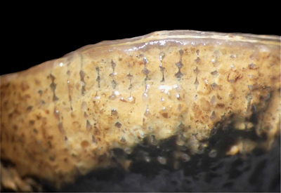
Repeated
binocular observations of epithecal growth wrinkles on the lower surface of Montlivaltia specimens indicate that their distribution is
not random but reflects rhythmic growth patterns. Although irregularities do
occur (Fig. 7 ![]() ) and can sometimes complicate age determination, it is unlikely
that the observed regularities result from random growth disturbances (Figs. 8
) and can sometimes complicate age determination, it is unlikely
that the observed regularities result from random growth disturbances (Figs. 8 ![]() - 9
- 9 ![]() - 10
- 10 ![]() ). The periodic signal can, however, be disrupted by non-periodic events,
such as the tilting of a coral within the substrate (Fig. 7
). The periodic signal can, however, be disrupted by non-periodic events,
such as the tilting of a coral within the substrate (Fig. 7 ![]() ).
).
|
|
Figure 7: Lateral
view of Montlivaltia Ech 2024-038 (originally identified as M.
decipiens,
Fontoy). Note the fairly regular annual banding and the growth wedge
(white arrow), reflecting an upright reorientation of the skeleton after initial
settlement. Smaller cycles can be observed. |
|
|
Figure 8: Proximal view of Montlivaltia
specimen Ech 2024-034 (originally identified as M. delabechei), Les Gimeys
Farm, Sexey-aux-Forges, coll. Gardet.
Note that the skeleton from the first year(s) is not covered by epitheca. Regular
annual banding is visible over four successive years. |
|
|
Figure 9:
Proximal view of Montlivaltia
Ech 2024 0 33, originally identified as M.
labechei, Bathonian, Conflans, coll. Groth. |
|
|
Figure 10:
Epithecal growth rhythms on a Montlivaltia
specimen Ech 2024 0 33, originally identified as M. labechei,
Bathonian, Conflans, coll. Groth. Note the
imbrication between annual growth bands and lunar regular growth bands
(highlighted
by white lines). |
The most evident rhythm is expressed by growth increments generally ranging between 1.5 and 3 mm. These increments correspond to alternating phases of diametric expansion and contraction: expansion phases are represented by broad convex bands, whereas contraction phases are marked by narrower, deeper constriction lines. This fairly consistent increment size is interpreted as representing the annual growth band. Such regularity enables the estimation of the corallum's age even when certain annual records are poorly developed or irregular. Measurements are taken horizontally in discoid phenotypes and vertically in cylindroid forms.
The
earliest growth stages are often not covered by epithecal tissue but instead
display radiating radial elements (Fig. 8 ![]() ) over a small area, where growth
rhythms cannot be discerned. In some specimens, the outer surface is only
partially covered by epitheca, leaving radial elements visible corresponding
with phases of reduced calcification.
) over a small area, where growth
rhythms cannot be discerned. In some specimens, the outer surface is only
partially covered by epitheca, leaving radial elements visible corresponding
with phases of reduced calcification.
The most distinct banding, interpreted as annual, is itself composed of smaller, subordinate increments that are locally strikingly regular. These micro-increments measure approximately 160 μm, and 12-13 of them can generally be counted within a single annual band, insofar as the annual limits can be clearly defined.
Univariate
analyses of Bathonian Montlivaltia
from Lorraine reveal a homogeneous population, in which no species-level
distinction can be inferred from the measured parameters (Fig.
11 ![]() ). Both large
and small diameters display unimodal and symmetric distributions around their
respective means. In contrast, height exhibits an almost unimodal but strongly
asymmetric distribution, positively skewed, with only four specimens
significantly taller than the rest.
). Both large
and small diameters display unimodal and symmetric distributions around their
respective means. In contrast, height exhibits an almost unimodal but strongly
asymmetric distribution, positively skewed, with only four specimens
significantly taller than the rest.
The distribution of septal number (Ns) is approximately symmetric, but distinctly leptokurtic, and centred on a value very close to 96, the value predicted by the model of Milne-Edwards and Haime (1848), according to which septal insertion follows the formula 6S₁ + 6S₂ + 12S₃ + 24S₄ + 48S₅ = 96 septa. Septal density, trabecular density, and fossa length all approximate normal distributions.
The estimated age-distribution is broadly unimodal, though a few exceptionally old and tall specimens may support alternative interpretations. Its positive skewness resembles that observed in the height distribution.
|
|
Figure 11:
Univariate analyses of Montlivaltia
from the Bathonian of Lorraine. Letters refer to the dimensions of type
material of species initially identified by Gardet (1947) where
available. ca = caryophyllata, la
= delabechei, nu = numismalis, mu =mulleri, tr =
trochoides, wa = waterhousei. |
Several bivariate
analyses are presented in Figure 12 ![]() . The XY plot of large versus small diameter
shows that most Montlivaltia
specimens maintain an approximately circular outline, although a few individuals
exhibit varying degrees of ellipticity. This graph does not support any
subdivision of the population based on this parameter.
. The XY plot of large versus small diameter
shows that most Montlivaltia
specimens maintain an approximately circular outline, although a few individuals
exhibit varying degrees of ellipticity. This graph does not support any
subdivision of the population based on this parameter.
The XY plot of large diameter versus height indicates that most Montlivaltia display a low, flattened morphology, while some individuals show increased vertical growth, reflecting a wide range of ontogenetic trajectories. The plot of height as a function of age produces an elongated cloud of points, suggesting a roughly linear correlation (R² = 0.6). Exponential and logarithmic regressions yield lower coefficients of determination (R² < 0.5). The plot of number of septa (Ns) as a function of age shows no linear relation. After the age of 4 years most Montlivaltia reach their maximum number of septa (not far from 100) and keep it.
A
multivariate analysis was performed on a smaller subset of specimens; however,
the sample size remains sufficient to produce meaningful results. A Principal
Component Analysis (PCA) using the correlation option in PAST (to account for
parameters measured in different units) was conducted (Fig. 13 ![]() ). The scatter
plot is primarily structured by the influence of the number of septa on the
first principal component, whereas diameters and height mainly stretch the cloud
along the second component. In contrast, biological age contributes little to
the overall inertia of the point cloud. The scatter plot does not reveal any
clear subdivision within the population. Moreover, the geographic distribution
of localities does not satisfactorily explain the structure of the scatter plot,
as localities with numerous specimens (Conflans and Fontoy) show substantial
overlap.
). The scatter
plot is primarily structured by the influence of the number of septa on the
first principal component, whereas diameters and height mainly stretch the cloud
along the second component. In contrast, biological age contributes little to
the overall inertia of the point cloud. The scatter plot does not reveal any
clear subdivision within the population. Moreover, the geographic distribution
of localities does not satisfactorily explain the structure of the scatter plot,
as localities with numerous specimens (Conflans and Fontoy) show substantial
overlap.
|
|
Figure 12:
Bivariate
analyses of Montlivaltia
from the Bathonian of Lorraine. Lettered data points correspond to Gardet's
(1947) initial species identifications (mu = muelleri, tr = trochoides). |
|
|
Figure 13:
Multivariate
analyses of Montlivaltia from
the Bathonian of Lorraine. |
A statistically homogeneous population does not necessarily correspond to a biologically homogeneous one. Overall, based on the combined results of univariate, bivariate, and multivariate analyses, the distribution of parameters appears relatively uniform, supporting the interpretation that the studied Montlivaltia specimens belong to a single species. However, several issues require careful consideration.
It is clear that the genus Montlivaltia has been excessively split in past taxonomic treatments. Little justification can be found for the numerous species names proposed by Gardet (1947) and other authors. Gardet (1947) did not justify his identifications, but it is assumed that he followed the traditional practice of distinguishing species based on diameter, number of septa and height as was commonly done by earlier authors (see for instance Fromentel & Ferry, 1865-1869). I, nevertheless, considered the authors who proposed alternative criteria for species discrimination (e.g., new parameters introduced by Alloiteau, 1958, or new types of graphical analyses proposed by Gill and Lafuste, 1971). However, as already concluded by Pandey and Fürsich (2003, p. 34) after their detailed analysis of character variation, all of these additional parameters are subject to substantial intraspecific variability. A recent population study of Middle Jurassic Montlivaltia from Tibet by Zhu et al. (2025) reached a similar conclusion, recognizing a single, morphologically variable species. Unfortunately, those authors assigned their population to the junior synonym Montlivaltia zangbeiensis Liao and Xia, 1985, which closely resembles older nominal species described from Europe [*].
(1) In the same publication, Zhu et al. (2025) also misassigned a species of Adelocoenia Orbigny, 1849, to the obsolete genus Pseudocoenia Orbigny, 1850 (see Lathuilière et al., 2020).
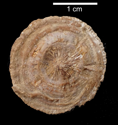
In
cases of broad intraspecific variability encompassing several previously named
taxa, interpreting specimens at the extremes of the variation range becomes
problematic. The unique Kobyphyllia
specimen (Fig. 14 ![]() ) is of particular interest: it occupies a central position
within the quantitative distribution but differs from the others only in
possessing a lamellar columella - a qualitative feature. The hypothesis that Kobyphyllia
represents merely an individual variant of Montlivaltia
is defensible, although no practical test currently allows verification of this
idea.
) is of particular interest: it occupies a central position
within the quantitative distribution but differs from the others only in
possessing a lamellar columella - a qualitative feature. The hypothesis that Kobyphyllia
represents merely an individual variant of Montlivaltia
is defensible, although no practical test currently allows verification of this
idea.
|
|
Figure 14:
Specimen
MAN 2024 0 44-24 here identified as a Kobyphyllia
because of its lamellar columella. |
At the end of the distribution, I am not entirely convinced that the
specimens originally designated as Montlivaltia
mülleri Koby, 1884, - characterized by a markedly tall growth form -
belong to the same biological unit. It remains uncertain whether this distinct
morphology reflects random variation, an ecophenotypic response, or a true
specific difference. This doubt is reinforced by septal microarchitectural
observations that are atypical for Montlivaltia.
These specimens are both taller and older than the others (Fig. 12 ![]() ) but could
not be included in all analyses because trabecular density and fossa length
could not be measured. An ecophenotypic interpretation is plausible: prolonged
growth may have been facilitated by stable soft substrates and appropriate
sedimentation rates. Notably, all previously named "M. mülleri"
(normally corrected into muelleri) specimens come from distinct outcrops (Gruyère and
Beney)
where they represent the only species of the genus present. Interestingly, even Goldfuss
(1829), despite the typological framework of his time, illustrated Montlivaltia
decipiens (then classified as Anthophyllum)
with both discoid and cylindroid morphologies. Much later, Gill and Lafuste
(1971) also proposed an ecophenotypic explanation. It is further possible that
the weak septal ornamentation observed in older specimens reflects biological
aging, involving the addition of lamellar layers that obscure the sharp relief
of younger trabeculae.
) but could
not be included in all analyses because trabecular density and fossa length
could not be measured. An ecophenotypic interpretation is plausible: prolonged
growth may have been facilitated by stable soft substrates and appropriate
sedimentation rates. Notably, all previously named "M. mülleri"
(normally corrected into muelleri) specimens come from distinct outcrops (Gruyère and
Beney)
where they represent the only species of the genus present. Interestingly, even Goldfuss
(1829), despite the typological framework of his time, illustrated Montlivaltia
decipiens (then classified as Anthophyllum)
with both discoid and cylindroid morphologies. Much later, Gill and Lafuste
(1971) also proposed an ecophenotypic explanation. It is further possible that
the weak septal ornamentation observed in older specimens reflects biological
aging, involving the addition of lamellar layers that obscure the sharp relief
of younger trabeculae.
Growth rhythms in corals may arise from various mechanisms. Some are internally regulated, reflecting compromises between the growth of soft tissues and that of the skeleton. Gill (1982) elegantly illustrated such patterns in the alternation in levels of pennulae in pennular corals, of auriculae in Stylinidae, and tabular development within and outside corallites in plocoid stylinids. In the present material, coordination between soft-tissue expansion and skeletal accretion was probably mediated by numerous vesicular dissepiments, enabling steady growth along the distal margins of the septa.
Other rhythmic patterns may record variations in growth rate linked to fluctuating environmental conditions. Since the pioneering work of Ma (1933, 1934, 1937), who emphasized seasonal influences on coral growth and their palaeontological value, many studies have identified annual banding on the epitheca or within the skeleton. The annual nature of these bands has often been confirmed through geochemical analyses in Recent corals, forming the basis of sclerochronology - a key approach in palaeoclimatic reconstruction (for recent reviews, see Helmle & Dodge, 2011; Peharda et al., 2021). The studied population of Bathonian Montlivaltia is situated at a rather high palaeolatitude (not far from 30° north), which was rather favourable for the record of seasonal contrasts.
Less currently, subordinate rhythms related to lunar or circadian cycles have also been reported. Their attribution to specific periodicity is not always convincing (see for example the so-called daily growth of the Recent Madracis Milne Edwards & Haime, 1849b, Florida in Wells (1970: Fig. 4) with growth increments of more than 80 micro-meters, which seem good candidates for synodic increments. Annual increments of 36-69 mm per year, calculated (and not observed) from supposed daily nested in monthly ones, for Carboniferous rugosans by Johnson and Nudds (1975) seem also very high. Scrutton (1998) has already pointed that these results are hardly compatible with recorded growth rates in rugosan corals. Scrutton (1970) mentioned that daily increments are generally thinner than 50 micrometers. Lunar periodicity was described by Scrutton (1970) who described Devonian corals, a period in which there was 399 days in a year, and, according to the Scrutton's data, around 31 days in a lunar cycle. Today there are 12.4 synodic months in a year and 29.5 days per month. Lunar cycles were reported in Lophelia pertusa (Linnaeus, 1758), a Recent non-zooxanthellate coral (a personal communication of S.A. Wainwright reported by Scrutton (1965), and by other authors in modern zooxanthellate taxa (Buddemeier, 1974; Weber et al., 1975; Chevalier, 1987). The fine-scale increments observed within the annual growth bands of Bathonian Montlivaltia are interpreted as lunar in origin, based on their numerical ratio with annual bands. I see no alternative explanation and infer, as a first hypothesis, that spring-neap tidal cycles may have influenced calcification rhythms in Bathonian from Lorraine. Tidal cycles have multiple effects on environmental factors that can vary significantly in intensity according to topographical conditions. If tidal currents are channelized, they can move the water masses down to the bathyal environment (Lambert & Roux, 1991), improving, for instance, oxygenation of the sea bottom, which is important for the calcification process. But lunar effects on reproductive cycles are also well established (Babcock et al., 1986; Baird et al., 2009) and widely known through the spectacular images of mass spawning events, which occur often during specific nights each year (Mera et al., 2025), and thus, cannot account for a regular monthly signal. Lin et al. (2021) have demonstrated that the moonrise light is synchronised with spawning in Dipsastraea speciosa (Dana, 1846). Eighty-percent of Recent corals are broadcasting spawner (Baird et al., 2021) but the remaining brooders are also influenced by lunar cycles (light or tides). Atoda (1947a; 1947b; 1951a; 1951b; 1951c) has observed regular monthly production of planula larvae for the brooding coral Pocillopora damicornis (Linnaeus, 1758), Stylophora pistillata (Esper, 1792), Seriatopora hystrix Dana, 1846, Acropora brueggemanni (Brook, 1891), Galaxea aspera Quelch, 1886. Then, the hypothesis of Montlivaltia caryophyllata being a brooding species with a monthly-paced planulation controlled by an internal mechanism synchronized on astronomical rhythms is plausible as well as the direct triggering of monthly growth ridges driven by external environmental controls.
Circadian rhythms have been attributed to diurnal expansion and contraction movements of soft tissues at the colony margins (Barnes, 1972; Beauvais & Chevalier, 1980). Such daily cycles are rarely preserved in fossils, though examples are reported from Paleozoic corals [Wells, 1963; Johnson & Nudds, 1975 (but daily increments?); Guillaume & Semenoff-Tian-Chansky, 1991]. In Recent corals, daily increments (≈365 per year) have even been used to estimate the deceleration of Earth's rotation since the Devonian (Wells, 1963, 1970) despite their problematic accuracy (Scrutton & Hipkin, 1973). No such microincrements were detected in studied samples, likely due to the diagenetic replacement of aragonite by calcite, which increased the size of crystals and obscured very fine structures in Mesozoic corals, at least in their usual state of preservation. Assuming an annual growth increment of 1.5 mm and a Jurassic year of 380 days, the expected daily growth increments would be approximately 4 μm.
Frankowiak et al. (2016) demonstrated that fine-scale banding within thickening deposits is regular in zooxanthellate corals and irregular in azooxanthellate ones. Unfortunately, this approach cannot be applied to the studied material, because the aragonite-calcite transformation has degraded fine-scale textures. The regularity of larger annual growth bands has also been proposed as an indicator of photosymbiosis (Stanley & Helmle, 2010). This observation supports the interpretation of Montlivaltia as a zooxanthellate coral, although the criterion requires further validation through statistical and actualistic data (see Caroselli et al., 2017, for slow, regular growth in Caryophyllia Lamarck, 1801.
Annual linear growth rates have also been used to distinguish photosymbiotic from non-photosymbiotic corals, with the former generally showing higher calcification efficiency. The Bathonian Montlivaltia growth rates fall within the range common to both categories. However, comparison with Kimmeridgian descendants offers additional insight. In Montlivaltia nattheimensis Milaschewitch, 1876, illustrated by Milaschewitch (1876: Pl. 44, fig. 2), an annual linear growth of 4.6 mm can be estimated - relatively high compared with solitary, non-zooxanthellate corals (Caryophyllia: 1.05-2.94 mm, Chevalier, 1987; Leptopsammia pruvoti Lacaze-Duthiers, 1897: <1 mm, Caroselli et al., 2012).
Corals, like trees, are sometimes described as virtually immortal. Indeed, very old colonies do exist. Chevalier (1987, p. 540) reported a Platygyra Ehrenberg, 1834, colony 500 years old in the Red Sea and a Pavona frondifera (Lamarck, 1816) measuring 44.5 × 24.6 m from Shikoku Island, noting that such large colonies are composite structures regenerated by peripheral planula settlement. More recently, Cuff (2024) documented a Pavona clavus (Dana, 1846) colony estimated at 300 years and 32-34 m in diameter.
In colonial corals, growth generally decreases with age (Chevalier, 1987). In the zooxanthellate coral Manicina areolata (Linnaeus, 1758), T.F. Goreau and N.I. Goreau (1960) demonstrated that calcification rate is inversely related to body weight (and thus size), whereas in specimens reared in darkness, growth rate remains constant. Despite the fact that both species share the trait of initial attachment to the substrate, later breaking free to live unattached within soft sediment, this statement does not parallel my observations in Montlivaltia, where early growth increments cover a smaller surface area.
It remains unclear whether age in these corals is biologically constrained or reflects an adaptive response to unstable, soft-bottom environments - possibly explaining their relatively short lifespans.
The
regenerative capacity of corals is well known, and recent studies have begun to
elucidate its mechanisms, including the discovery of adult stem cells in the
colonial coral Stylophora pistillata (Levanoni et al.,
2024). The mean age of Bathonian Montlivaltia
population under study is six years, with the oldest specimen reaching 22 years.
This individual, initially identified as M.
mülleri, achieved its tall form through extended growth. Statistically, age
(inferred from growth bands) correlates with height (Fig. 12 ![]() ). With a mean lifespan of six years, these corals are, of course, far from
immortal and much shorter-lived than large colonial forms. Whether their death
was biologically programmed or environmentally induced remains uncertain.
). With a mean lifespan of six years, these corals are, of course, far from
immortal and much shorter-lived than large colonial forms. Whether their death
was biologically programmed or environmentally induced remains uncertain.
Finally, age estimation in fossils is of particular interest when exploring relationships between ontogeny and phylogeny in a lineage. In palaeontology, size is often used as a proxy for age - an assumption that can lead to confusion between dwarfism and progenesis, or between gigantism and hypermorphosis (Alberch et al., 1979; Dommergues et al., 1986). Direct age estimation, thus, provides an additional parameter for understanding size variation in the evolutionary history of the genus Montlivaltia. Population-based approaches will remain essential to document this lineage further.
The studied corals of the solitary genus Montlivaltia from the Bathonian of Lorraine appear to represent a single homogenous population, with only minor, possibly disputable exceptions at the extremes of the variation range.
Their epitheca displays regular growth bands organized at two hierarchical levels; the interleaving of these levels suggests control by solar and lunar cycles corresponding to annual and synodic (lunar) periodicities.
Consequently, the biological age of these Montlivaltia specimens can be determined: the mean age is relatively low (≈6 years), while the oldest individual observed reached 23 years.
Age determination opens a window for studying lineages and for a better understanding of evolutionary processes within the genus and comparable taxa.
The regularity of growth further supports the hypothesis that this genus was photosymbiotic.
It is a pleasure to thank Thibaut Keinerknecht for the work he did to help me in the MAN collections (Nancy) and Markus Aretz for helping me with the Palaeozoic literature. Many thanks also to Thomas Brachert and an anonymous reviewer for their constructive and helpful comments.
Alberch P., Gould S.J., Oster G.F. & Wake D.B. (1979).- Size and shape in ontogeny and phylogeny.- Paleobiology, vol. 5, no. 3, p. 296-317.
Alloiteau J. (1957).- Contribution à la systématique des madréporaires fossiles.- Éditions CNRS, Paris, 462 p.
Alloiteau J. (1958).- Monographie des madréporaires fossiles de Madagascar.- Annales géologiques de Madagascar, Antananarivo, no. XXV, 218 p.
Atoda K. (1947a).- The larva and post-larval development of some reef-building corals. I. Pocillopora damicornis.- Science Reports of the Tôhoku Imperial University (4th serie, Biology), Sendai, vol. XVIII, no. 1, p. 24-47.
Atoda K. (1947b).- The larva and post-larval development of some reef-building corals. II. Stylophora pistillata (Esper).- Science Reports of the Tôhoku Imperial University (4th serie, Biology), Sendai, vol. XVIII, no. 1, p. 48-64.
Atoda K. (1951a).- The larva and post-larval development of some reef-building corals. V. Seriatopora hystrix Dana.- Science Reports of the Tôhoku Imperial University (4th serie, Biology), Sendai, vol. XIX, no. 1, p. 33-39.
Atoda K. (1951b).- The larva and post-larval development of the reef-building corals. III. Acropora brüggemanni (Brook).- Journal of Morphology, vol. 89, no. 1, p. 1-15.
Atoda K. (1951c).- The larva and post-larval development of the reef-building corals. IV. Galaxea aspera Quelch.- Journal of Morphology, vol. 89, no. 1, p. 17-35.
Babcock R., Bull G., Harrison P., Heyward A., Oliver J., Wallace C. & Willis B. (1986).- Synchronous spawnings of 105 scleractinian coral species on the Great Barrier Reef.- Marine Biology, vol. 90, p. 379-394.
Baird A.H., Guest J.R. & Willis B. (2009).- Systematic and biogeographical patterns in the reproductive biology of scleractinian corals.- Annual Review of Ecology, Evolution, and Systematics, San Mateo - CA, vol. 40, p. 551-571.
Barnes D.J. (1972).- The structure and formation of growth-ridges in scleractinian coral skeletons.- The Royal Society of London, Proceedings B, vol. 182, no. 1068. p. 331-350.
Baron-Szabo R.C. & Fernandez-Mendiola P.A. (1997).- Cretaceous scleractinian corals from the Albian of Cabo de Ajo (Cantabria Province, N-Spain).- Paläontologische Zeitschrift, vol. 71, p. 35-50.
Beauvais L. & Chevalier J.P. (1980).- La croissance périodique chez les scléractiniaires actuels et fossiles.- Bulletin de la Société zoologique de France, Paris, t. 105, no. 2, p. 301-308.
Brook G. (1891).- LIV.- Descriptions of new species of Madrepora in the collections of the British Museum.- Annals and Magazine of Natural History (Series 6), London, vol. 8, no. 48, p. 458-471.
Buddemeier R.W. (1974).- Environmental controls over annual and lunar monthly cycles in hermatypic coral calcification. In: Proceedings of the Great Barrier Reef Committee.- Great Barrier Reef Committee, Brisbane, p. 259-267.
Caroselli E., Zaccanti F., Mattioli G., Falini G., Levy O., Dubinsky Z. & Goffredo S. (2012).- Growth and demography of the solitary scleractinian coral Leptopsammia pruvoti along a sea surface temperature gradient in the Mediterranean Sea.- PLOS One, San Francisco - CA, vol. 7, no. 6, article e37848, 10 p. DOI: 10.1371/journal.pone.0037848
Caroselli E., Ricci F., Brambilla V., Marchini C., Tortorelli G., Airi V., Mattioli G., Levy O., Falini G., Dubinsky Z. & Goffredo S. (2017).- Growth, population dynamics, and reproductive output model of the non-zooxanthellate temperate solitary coral Caryophyllia inornata (Scleractinia, Caryophylliidae).- Limnology and Oceanography, vol. 62, no. 3, p. 1111-1121. DOI: 10.1002/lno.10489
Chevalier J.-P. (1987).- Ordre des Scléractiniaires. In: Grassé P.P. (ed.), Traité de Zoologie, vol. 3, fasc. 3.- Masson, Paris, p. 403-764.
Cuff M. (2024).- World's largest coral is 300 years old and was discovered by accident.- New Scientist, New York - NY, no. 3518, p. 18
Dana J.D. (1846-1849).- Zoophytes. United States Exploring Expedition during the years 1838-1842.- Lea and Blanchard, Philadelphia - PA, 740 p. (1846: p. 1-120, p. 709-720; 1848: p. 121-708, p. 721-740; 1849: atlas 61 Pls.).
Dommergues J.-L., David B. & Marchand D. (1986).- Les relations ontogenèse-phylogenèse : Applications paléontologiques.- Geobios, Villeurbanne, vol. 1, no. 3, p. 335-356.
Ehrenberg C.G. (1834).- Beiträge zur physiologischen Kenntniss der Corallenthiere im allgemeinen, und besonders des rothen Meeres, nebst einem Versuche zur physiologischen Systematik derselben.- Abhandlungen der Königlichen Akademie der Wissenschaften, Berlin, Jahre 1832, Erster Theil, p. 225-380. URL: https://www.biodiversitylibrary.org/page/29725862
Esper E.J.C. (1788-1830).- Die Pflanzenthiere in Abbildungen nach der Natur mit Farben erleuchtet nebst Beschreibungen.- Raspischen Buchhandlung, Nuremberg: 3 vols. text, 2 vols. Pls. DOI: 10.11588/diglit.2885
Frankowiak K., Kret S., Mazur M., Meibom A., Kitahara M. & Stolarski J. (2016).- Fine-scale skeletal banding can distinguish symbiotic from asymbiotic species among modern and fossil scleractinian corals.- PLOS One, San Francisco - CA, vol. 11, no. 1, article e0147066, 21 p. DOI: 10.1371/journal.pone.0147066
Fromentel É. de & Ferry H. de (1865-1869).- Paléontologie Française ou Description des fossiles de la France T. XII, Zoophytes. 5ème classe. Coralliaires : 1ère série: Animaux invertébrés : Terrain Jurassique.- Masson, Paris, 1865a: p. 1-48, Pls. 1-12; 1865b: p. 49-96, Pls. 13-24; 1866: p. 97-144, Pls. 25-36; 1867: p. 145-192, Pls. 37-48; 1869: p. 193-240, Pls. 49-60.
Gardet G. (1947).- Le Bathonien de la Lorraine.- Bulletin des Services de la Carte géologique de la France, Paris, vol. XLV, no. 217, p. 327-394.
Gill G.A. (1982).- A supposed rhythmic mechanical process in coral skeletal growth. In: Montanaro-Gallitelli E. (ed.), Palaeontology, essential of historical geology.- S.T.E.M. Mucchi, Modena, p. 445-466.
Gill G.A. & Lafuste J.G. (1971).- Madréporaires simples du Dogger d'Afghanistan : Étude sur les structures de type Montlivaltia.- Mémoires de la Société géologique de France (Nouvelle Série), Paris, t. L, no. 115, 40 p.
Goldfuss A. (1829).- Petrefacta Germaniæ tam ea, quae in Museo Universitatis Regiae Borussicae Fridericiae Wilhelmiae Rhenanae servantur quam alia quaecunque in Museis Hoeninghusiano Muensteriano aliisque extant, iconibus et descriptionibus illustrata.- Erster Theil, Arnz & Cie., Düsseldorf, 252 p. (LXXI Pls.).
Goreau T.F. & Goreau N.I. (1960).- Physiology of skeleton formation in corals. III. Calcification rate as a function of colony weight and total nitrogen content in the reef coral Manicina areolata (Linnaeus).- Biological Bulletin, Chicago - IL, vol. 118, p. 419-429.
Grottoli A. (2001).- Past climate from corals. In: Steele J., Thorpe S. & Turekian K. (eds.), Encyclopedia of ocean sciences.- Academic Press, London, p. 2098-2107.
Guillaume M. & Semenoff-Tian-Chansky P. (1991).- Stries semi-journalières chez un Tétracoralliaire (Bothrophyllum proteum) du Carbonifère supérieur. Implication dans la détermination des rythmes de croissance.- Comptes Rendus de l'Académie des Sciences (série II), Paris, t. 312, p. 1401-1407.
Helmle K.P. & Dodge R.E. (2011).- Sclerochronology. In: Hopley D. (ed.), Encyclopedia of modern coral reefs: Structure, form and process.- Springer, Berlin, p. 958-966.
Johnson G.A.L. & Nudds J.R. (1975).- Carboniferous coral geochronometers. In: Rosenberg G.D. & Runcorn S.K. (eds.), Growth rhythms and the history of the Earth's rotation.- Wiley, Oxford, p. 27-41.
Koby F. (1884).- Monographie des polypiers jurassiques de la Suisse (3).- Mémoires de la Société Paléontologique Suisse, Genève, vol. X, p. 109-148.
Koby F. (1889).- Monographie des polypiers jurassiques de la Suisse (9).- Mémoires de la Société Paléontologique Suisse, Genève, vol. XVI, p. 457-586.
Lacaze-Duthiers H. (1897).- Faune du Golfe de Lion. Coralliaires. Zoanthaires sclérodermés (Deuxième mémoire).- Archives de Zoologie expérimentale et générale (3e Série), Paris, t. V, 249 p. URL: https://www.bnsp.insee.fr/ark:/12148/bpt6k5456995s
Lamarck J.B. de (1801).- Systême des animaux sans vertèbres, ou tableau général des classes, des ordres et des genres de ces animaux.- Chez l'auteur, Verdière, Paris, viii + 432 p. URL: https://gallica.bnf.fr/ark:/12148/bpt6k5552124z
Lamarck J.B. de (1816).- Histoire naturelle des animaux sans vertèbres. Tome second.- Verdière, Paris, 566 p. URL: https://www.biodiversitylibrary.org/page/13298812
Lambert B. & Roux M. (1991).- L'environnement carbonaté bathyal en Nouvelle-Calédonie (programme envimarges).- Documents et Travaux IGAL, Cergy-Pontoise, no. 15, 213 p.
Lamouroux J. (1821).- Exposition méthodique des genres de l'ordre des polypiers.- Mme veuve Agasse, Paris, 115 p. URL: https://www.biodiversitylibrary.org/page/11721203
Lathuilière B., Baron-Szabo R.C., Charbonnier S. & Pacaud J.M. (2020).- The Mesozoic scleractinian genus Adelocoenia (Stylinidae) and its Jurassic species.- Carnets Geol., vol. 20, no. 19, p. 367-406. DOI: 10.2110/carnets.2020.2019
Lathuilière B. & Michel B. (2019).- A new Bathonian coral fauna in Lorraine (Middle Jurassic, France). In: Bosellini F., Aretz M., Papazzoni C.A. & Vescogni A. (eds.), 13th International Symposium on Fossil Cnidaria and Porifera 2019, Modena, Italy. Abstract Book.- Unimore, Modena, p. 38.
Levanoni J., Rosner A., Lapidot Z., Paz G. & Rinkevich B. (2024).- Coral tissue regeneration and growth is associated with the presence of stem-like cells.- Journal of Marine Science and Engineering, Basel, vol. 12, article 343, 20 p.
Liao W.-H. & Xia J.-B. (1985).- Upper Jurassic and lower Cretaceous Scleractinia from Bangoin district of northern Xizang (Tibet).- Memoirs of the Nanjing Institut of Geology and Paleontology, vol. 21, p. 119-178.
Lin C.-H., Takahashi S., Mulla A.J. & Nozawa Y. (2021).- Moonrise timing is key for synchronized spawning in coral Dipsastraea speciosa.- PNAS, Washington - DC, vol. 118, no. 34, 6 p. DOI: 10.1073/pnas.2101985118
Linnaeus C. (1758).- Systema Naturæ per regna tria naturæ, secundum classes, ordines, genera, species, cum characteribus, differentiis, synonymis, locis.- Holmiæ, Stockholm, t. I, 824 p. URL: https://www.biodiversitylibrary.org/page/726886
Ma T.Y.H. (1933).- 116. On the seasonal change of growth in some Palaeozoic corals.- Proceedings of the Imperial Academy, Sendai, vol. IX, no. 8, p. 407-409. DOI: 10.2183/pjab1912.9.407
Ma H.T.Y. (1934).- On the growth rate of reef corals and the sea water temperature in the Japanese Islands during the latest geological times.- Science Reports of the Tôhoku Imperial University, Sendai, vol. X, p. 165-189. URL: https://tohoku.repo.nii.ac.jp/records/10699
Ma H.T.Y. (1937).- On the growth rate of reef corals and its relation to sea water temperature.- Palaeontologia Sinica (series B), Beijing, vol. 16, fasc. 1, p. 1-426.
Mera H., Edwards A.J., Guest J.R., Lachs L., Quigley K.M., Willis B.L. & Baird A.H. (2025).- Patterns of coral spawning in the Palm Islands, Great Barrier Reef.- Coral Reefs, vol. 44, p. 1789-1803. DOI: 10.1007/s00338-025-02733-7
Milaschewitsch C. (1876).- Die Korallen der Nattheimer Schichten.- Palaeontographica, Band 21, Lieferung 8 (1875), p. 205-244.
Milne Edwards H. & Haime J. (1848).- Recherches sur les polypiers. Observations sur la structure et développement des polypiers en général.- Annales des Sciences naturelles (3e Série), Paris, t. IX, p. 37-89.
Milne Edwards H. & Haime J. (1849a).- Mémoire sur les Polypes appartenant à la famille des Oculinides, au groupe intermédiaire des Pseudastréides et à la famille des Fongides.- Comptes rendus hebdomadaires des Séances de l'Académie des Sciences, Paris, t. XXIX, p. 67-73. URL: https://gallica.bnf.fr/ark:/12148/bpt6k2986m
Milne Edwards H. & Haime J. (1849b).- Recherches sur les polypiers (4:) Monographie des Astréides (1:) Eusmiliens.- Annales des Sciences naturelles (3e Série), Paris, t. X, p. 209-320.
Orbigny A. d' (1849).- Note sur des polypiers fossiles.- Masson, Paris, 12 p.
Orbigny A. d' (1850).- Prodrome de paléontologie stratigraphique universelle des animaux mollusques et rayonnés.- Masson, Paris, 2e vol., 428 p. URL: https://www.biodiversitylibrary.org/page/41091877
Pandey D.K. & Fürsich F.T. (2003).- Jurassic corals of east-central Iran.- Beringeria, Würzburg, Heft 32, p. 3-138.
Peharda M., Schöne B.R., Black B.A. & Corrège T. (2021).- Advances of sclerochronology research in the last decade.- Palæogeography, Palæoclimatology, Palæoecology, vol. 570, article 110371, 16 p.
Quelch J.J. (1886).- The Voyage of H.M.S. Challenger. Zoology.- Report on the reef-corals collected by H.M.S. 'Challenger' during the years 1873-76, Edinburgh, vol. 16, part XLVI, 203 p. (XII Pls.). URL: https://www.biodiversitylibrary.org/page/12035017
Scrutton C.T. (1965).- Periodicity in Devonian coral growth.- Palaeontology, vol. 7, part 4, p. 552-558.
Scrutton C.T. (1970).- Evidence for a monthly periodicity in the growth of some corals. In: Runcorn S.K. (ed.), Palaeogeophysics.- Academic Press, London & New York, p. 11-16.
Scrutton C.T. (1998).- The Palaeozoic corals, II: Structure, variation and palaeoecology.- Proceedings of the Yorkshire geological Society, vol. 52, no. 1, p. 1-57.
Scrutton C.T. & Hipkin R.G. (1973).- Long-term changes in the rotation rate of the Earth.- Earth Science Reviews, vol. 9, no. 3, p. 259-274.
Stanley G.D. & Helmle K.P. (2010).- Middle Triassic coral growth bands and their implication for photosymbiosis.- Palaios, Lawrence - KS, vol. 25, p. 754-763.
Stolarski J. (1995).- Ontogenetic development of the thecal structures in caryophylline scleractinian corals.- Acta Palaeontologica Polonica, Warsaw, vol. 40, no. 1, p. 19-44.
Weber J.N., Deines P., White E.W. & Weber P.H. (1975).- Seasonal high and low density bands in reef coral skeletons.- Nature, London, vol. 255, p. 697-698.
Wells J.W. (1956).- Scleractinia. In: Moore R.C. (ed.), Treatise on invertebrate paleontology.- Geological Society of America, Boulder - CO; University of Kansas Press, Lawrence - KS, vol. F, p. 328-444.
Wells J.W. (1963).- Coral growth and geochronometry.- Nature, London, vol. 197, p. 948-950.
Wells J.W. (1970).- Problems of annual and daily growth rings in corals. In: Runcorn S.K. (ed.), Palaeogeophysics.- Academic Press, London & New York, p. 3-9.
Zany D. & Lathuilière B. (2018).- Bouvron (54) : Marnes du Bathonien - GAEC du Grand Parc.- Contribution ORAGE originale à la Banque de Données du Sous-Sol, Nancy, no. 16, 8 p. URL : https://orage.univ-lorraine.fr/s/orage/item/5605
Zhu X.P., Liang K., Liao W.-H., Yin J.R., Rao X. & Zhang Y.C. (2025).- Scleractinian corals from the Middle Jurassic Buqu Formation, Qiangtang block and their palaeogeographic implications.- Palaeoworld, vol. 35, no. 1, article 200978, 15 p.